Category: biophysics

Fast assessments of 3D shapes within cell populations

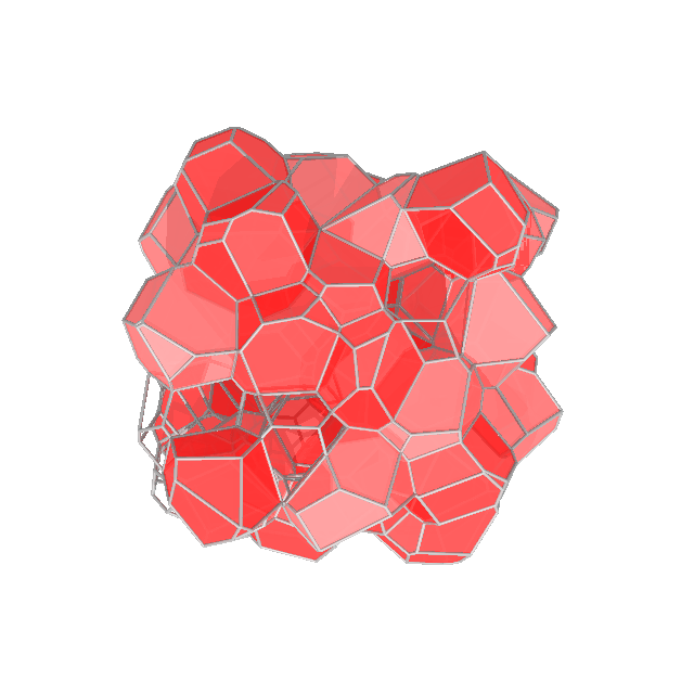
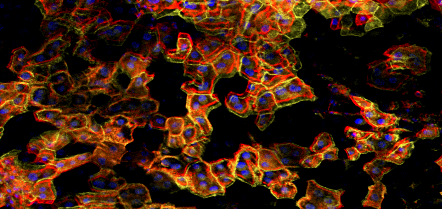
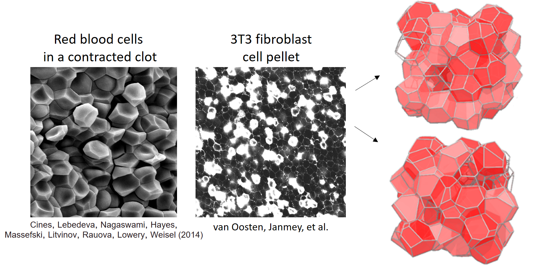
Within biological tissues, cell shape may be indicative of the chemical and mechanical micro-environment.  Experiments on isotropic and homogeneous packings of cells, as shown below, connect the 3D shapes with tissue properties.  Unfortunately, quantitatively measuring 3D shapes of cells is burdensome, requiring high-quality confocal microscopy and image post-processing.  Another approach to quantifying 3D cell shapes uses 2D imagery without ever reconstructing 3D shapes.  Instead, cell-vertex models with quasi realistic geometry can be used to  generate mappings between 3D shapes of cells and the ensemble of 2D shapes seen in a single image.   Researchers may now trace cells in simple 2D imagery to determine the distribution of shapes, and compare them to the model to determine the ensemble of 3D cell shapes.

We are working to release a software package to provide these 3D shape estimates from traces of 2D cells.  This method has allowed us to compare predictions from the 3D cell vertex model with experimental measurements.


Mechanics of biological tissue

The stiffness of a tissue reflects its cell-level properties. For instance, it is well known that increased stiffness can be an indicator a rapidly growing cancer in a tissue.  At UPenn, I am using simulations and theory, supported by experiments with the Penn Science of Oncology Center, to develop the biophysical understanding of tissue mechanics.

On short time scales, biological tissues have the mechanical properties of polymer networks embedded with cells and fluid. This is a complicated composite from a materials physics perspective. Experiments study simplified tissues, with cells alone with no extracellular matrix, or of polymer networks with embedded cell-like particles.  A long-term goal is to increase the utility of stiffness in characterizing the microscopic structure of the tissue.


We developed a 3D cell-vertex model based on the Voronoi tesselation to study the mechanical properties of confluent cell tissues. (TAS)
Modelling polymer networks embedded with beads helps interpret the experimental measurements of elastic moduli. (TAS)
Polymer matrix serves many mechanical roles in a tissue. In addition to providing mechanical support itself, it can also prevent cells from rearranging on long time scales. Simulations separate the two effects by choosing the topology of the fiber network. (TAS)
Confocal microscopy of liver tissue shows the changes in contact topology of the cells after straining. (TAS with LiKang Chin)

Read more at this page.