Fast assessments of 3D shapes within cell populations
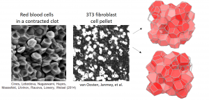
Within biological tissues, cell shape may be indicative of the chemical and mechanical micro-environment. Experiments on isotropic and homogeneous packings of cells, as shown below, connect the 3D shapes with tissue properties. Unfortunately, quantitatively measuring 3D shapes of cells is burdensome, requiring high-quality confocal microscopy and image post-processing. Another approach to quantifying 3D cell shapes uses 2D imagery without ever reconstructing 3D shapes. Instead, cell-vertex models with quasi realistic geometry can be used to generate mappings between 3D shapes of cells and the ensemble of 2D shapes seen in a single image. Researchers may now trace cells in simple 2D imagery to determine the distribution of shapes, and compare them to the model to determine the ensemble of 3D cell shapes.
We are working to release a software package to provide these 3D shape estimates from traces of 2D cells. This method has allowed us to compare predictions from the 3D cell vertex model with experimental measurements.

Ive read several just right stuff here Certainly price bookmarking for revisiting I wonder how a lot effort you place to create this kind of great informative website